
PurposePeriodontal ligament stem cells (PDLSCs) have appreciable potential to be used as a way of reaching periodontal regeneration resulting from their noteworthy proliferative properties and secretory features. In specific, PDLSCs secrete vascular endothelial growth factor (VEGF) which boosts angiogenesis and osteogenesis.
The ensuing restore and growth of blood vessels and onerous tissues which might happen within the presence of these cells could possibly be central to an efficient periodontal regeneration process.The bacterial biofilm of tooth floor associated to the periodontium may present both an inhibition or a stimulus to various factors concerned in a regenerative course of.
Cell tradition experiments have been investigated in vitro by including lipopolysaccharide (LPS) to the tradition medium however the impact of numerous focus of LPS in these circumstances has not been investigated. Therefore, this examine aimed to research the impact of LPS concentrations on proliferation of PDLSCs in vitro and on their secretion of VEGF.
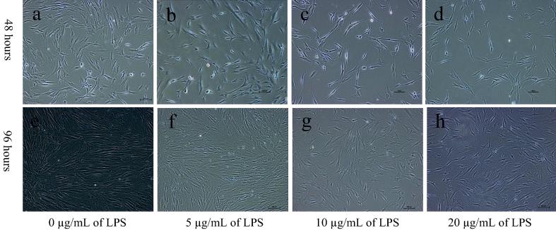
Materials and strategiesPDLSCs have been handled with 0, 5, 10 and 20 µg/mL of Escherichia coli LPS. At 48 and 96 h, whole cell numbers of management and LPS handled PDLSCs have been counted by haemocytometer underneath a microscope.
The VEGF focus within the conditioned media of the PDLSCs was measured by ELISA.Rate of cell proliferation of PDLSCs decreased considerably in all LPS handled teams at each 48 h and 96 h aside from the group handled with 5 µg/mL of LPS at 48 h. At each 48 and 96 h, VEGF secretion from PDLSCs was diminished considerably in any respect three LPS concentrations.
There was no statistically vital distinction in cell proliferation and the quantity of VEGF secretion of PDLSCs among the many teams handled with completely different LPS concentrations.
No statistically vital change was present in cell proliferation of LPS handled PDLSCs over time, whereas VEGF secretion of PDLSCs was discovered to have elevated considerably with time regardless of the LPS therapy.LPS diminished cell proliferation and VEGF secretion of PDLSCs, suggesting that periodontal pathogens may scale back the potential of PDLSCs in periodontal regeneration.
Yet, LPS handled PDLSCs remained viable and VEGF secretion elevated considerably over time. Further analysis is required to check the potential use of PDLSCs in periodontal regeneration and the connection of biofilm LPS accumulations.
Vascular Endothelial Growth Factor-Transfected Bone Marrow Mesenchymal Stem Cells Improve the Recovery of Motor and Sensory Functions of Rats With Spinal Cord Injury.
Basic science.The intention of this examine was to look at the impact of vascular endothelial growth factor (VEGF)-transfected bone marrow mesenchymal stem cells (BMSCs) on the restoration of motor and sensory features of rats with spinal wire harm (SCI).
There is not any efficient therapy to guard in opposition to SCI. BMSCs have been broadly utilized to the therapy of nervous system injury as a result of perform of immediate neurite growth and inhibition of demyelination following harm.
VEGF-transfected BMSCs have been injected to rats with SCI and the restoration of motor and sensory features was noticed. The Basso, Beattie, and Bresnahan, mechanical withdrawal threshold and thermal withdraw latency grading was performed to evaluate the restoration standing of motor and sensory features of the SCI rats. The expression of VEGF, CD31, and NF200 was detected by immunofluorescence.
The restoration of the rat motor and sensory features within the VEGF-transfected BMSC (BMSC-VEGF) group was increased than these of the opposite teams with the exception of the Sham group (P < 0.05). The expression of the CD31 and NF200 proteins within the rat SCI areas was the best within the BMSC-VEGF group, whereas the survival of BMSC within the BMSC-VEGF group was elevated in contrast with that within the BMSC-Ad group.
In addition, the injection of VEGF-transfected BMSCs can enhance the angiogenesis of the injured space and retain the survival of injected cells and neurons.The injection of BMSC-VEGF improved the restoration of motor perform in SCI rats.N/A.